Metabolism
on-line - the virtual tutorial room
copyright © 2008 - 2015 David A Bender
Poisoned by unripe ackee fruit
 HG gave a small dinner party, and in honour of one of the guests who was Jamaican,
prepared a traditional Jamaican dish, ackee with rice and salt fish, as well
as a number of other dishes.
HG gave a small dinner party, and in honour of one of the guests who was Jamaican,
prepared a traditional Jamaican dish, ackee with rice and salt fish, as well
as a number of other dishes.
Unfortunately, she had used unripe ackee fruit.
Only two of the guests ate any of ackee dish, and about an hour after the meal both became unconscious. An ambulance was called and they were taken to hospital. Emergency measurement of their blood glucose showed that it was dangerously low: 1.9 mmol /L in one case and 2.1 mmol /L in the other.
What is the normal range of blood glucose?
What emergency treatment should they received?
The normal range of blood glucose is 3.5 - 5.5 mmol /L,
but may rise to 8 mmol /L after a meal.
The obvious emergency treatment would be intravenous glucose
Why does a very low blood glucose concentration lead to loss of consciousness?
The brain is more or less completely reliant on glucose as its metabolic fuel, except in prolonged starvation, and the brain accounts for about 20% of whole body resting energy expenditure. Most of this energy expenditure is involved in transporting sodium and potassium ions across nerve membranes, against their concentration gradient, to maintain electrical activity. The sodium pump (in the brain and all other tissues) accounts for about 22% of whole body resting energy expenditure.
How can sodium and potassium ions be transported across cell membranes against their concentration gradient?
To transport ions against their concentration gradient requires an input of energy.
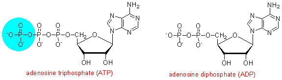
Here we need to introduce the compound adenosine triphosphate (ATP), which has a high free energy of hydrolysis to yield adenosine disphosphate (ADP) and inorganic phosphate. In addition to its role in ion transport, you will see later in this exercise how ATP can be used to drive endothermic reactions in the thermodynamically unfavoured direction, and how it is involved in muscle contraction and hence the performance of physical work.
You will see in later exercises how the phosphorylation of ADP back to ATP is linked to the oxidation of metabolic fuels, so that it acts as an intermediate between the oxidative pathways that are energy yielding and the various processes that are energy requiring.
What happens in sodium / potassium transport is that the carrier in the membrane can exist in two conformations:
With ATP bound it faces inwards, and binds 3 x sodium ions. The binding of sodium ions leads to phosphorylation of the carrier, release of ADP and a conformational switch so that it now faces outwards and expels sodium ions into the extracellullar fluid.
Two potassium ions now bind to the outward facing transporter, causing dephosphorylation. ATP binds to the dephosphorylated transporter, causing it to flip to face inwards again, expelling potassium ions into the cytosol, and ready to bind sodium ions again.
Click here to see more detail of the steps in sodium / potassium transport
Click here to see an animation of this. (Depending on your browser, you may be able to run this animation directly, or it may be downloaded and saved as p-type.exe)
How can ATP be used to drive endothermic reactions in the thermodynamically unfavoured direction?
An endothermic reaction requires an input of energy if it is to proceed in the thermodynamically unfavoured direction. In the laboratory this is often achieved by heating the reaction mixture (often to a very high temperature), something that is obviously not possible in the body. There are two ways in which ATP can be used - both result overall in the hydrolysis of ATP to ADP and inorganic phosphate, which is a highly exothermic reaction:
Phosphorylation of the enzyme as an intermediate stage of the reaction, so creating a charged group at the active site of the enzyme which aids catalysis (in a later exercise you will see how enzymes catalyse reactions by lowering the activation energy)
Phosphorylation of the substrate, so creating a group that can readily be displaced.

How is ATP involved in muscle contraction?
The key proteins in muscle are myosin, a fibrous protein that has ATPase activity in the light chains that make up the head region of the protein, and actin, a globular protein. Molecules of actin are arranged along a filament of tropomyosin (interspersed with molecules of troponin, a calcium-binding regulatory protein). The head regions of myosin are associated with actin, and the process of contraction involves the head regions moving along the actin chain to become associated with another actin molecule, and the tail regions sliding over one another.
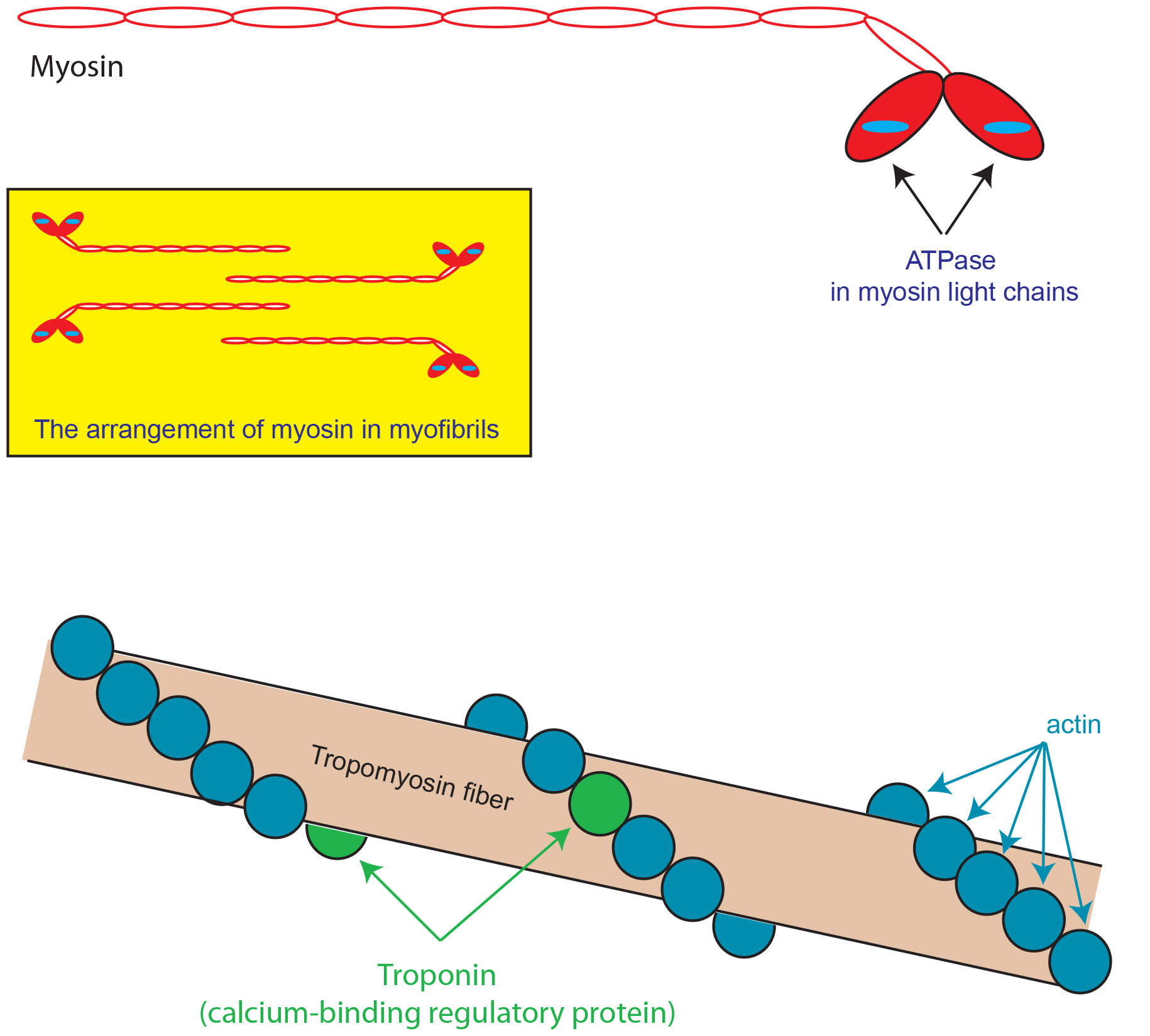
Myosin that is tightly bound to actin has ADP bound. Replacement of this ADP by ATP loosens the binding to actin; hydrolysis of the ATP to ADP and phosphate loosens it further, and release of phosphate (leaving ADP bound to myosin) causes the myosin fibres to slide over each other and the head regions to be associated with actin molecules further along the chain.
Click here to see more detail of the steps in muscle contraction
Click here to see an animation of muscle contraction. (Depending on your browser, you may be able to run this animation directly, or it may be downloaded and saved as p-type.exe)
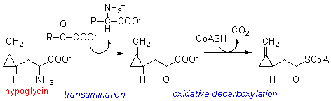
Unripe
ackee contains an unusual amino acid, called hypoglycin, because of its effect
in causing (potentially fatal) hypoglycaemia. The amount of hypoglycin decreases
as the fruit ripens, so that ripe ackee is not toxic. Hypoglycin is metabolised
to a coenzyme A derivative that does not undergo further metabolism.