Metabolism
on-line - the virtual tutorial room
copyright © 2008 - 2015 David A Bender
Alanine released from muscle in fasting
Wahren and coworkers reported a series of studies in which catheters were inserted into a brachial artery, a femoral vein and a hepatic vein (via an antecubital vein) in fasting volunteers. This meant that they could withdraw blood samples from three sites simultaneously to measure arterio-venous differences in concentrations of metabolites across the leg muscles (arterio-femoral vein difference) and across the liver (arterio-hepatic vein difference).
The arterio-venous difference is the concentration of the metabolite in arterial blood minus the concentration in the venous blood.
A negative a-v difference represents output of the metabolite by the tissue
A positive a-v difference represents uptake of the metabolite by the tissue
glucose |
lactate |
|
| arterial - femoral vein difference, mmol /L | 0.19 ± 0.02 |
-0.14 ± 0.03 |
| arterial - hepatic vein difference, mmol /L | -0.80 ± 0.08 |
0.21 ± 0.02 |
(From data reported by Wahren J. et al Journal of Clinical Investigation 51: 1870 1972)
What conclusions can you draw from these results?
Even in the fasting state there is a small uptake of glucose by muscle, but much of the glucose uptake will presumably have come from red blood cells, as will the output of lactate.
There is a considerable output of glucose by the liver. Some of this will come from liver glycogen, but much will come from gluconeogenesis after an overnight fast.
What are the likely substrates for gluconeogenesis in liver in the fasting state?
There will be a small amount of lactate, mainly from red blood cells, although resting muscle takes up glucose and metabolises it to some extent anaerobically, putting out lactate. However, the main substrates for gluconeogenesis in the fasting state are amino acids.
The diagram shows the arterial - femoral vein differences in concentrations of amino acids
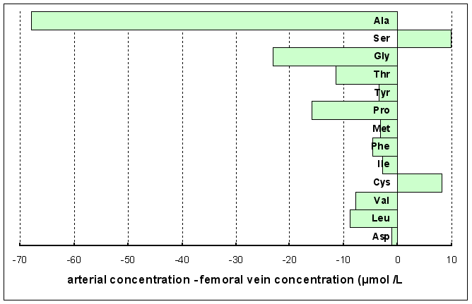
(From data reported by Wahren J. et al Journal of Clinical Investigation 51: 1870 1972)
What conclusions can you draw from these results?
In most cases the arterio-venous difference is negative, showing that there is an output of most amino acids form muscle in the fasting state, although it takes up moderate amounts of serine and cysteine.
The output of alanine is 3-fold higher than that of any other amino acid, amounting to ~60% of the total amino acid output.
Where have these amino acids come from?
They must have come from catabolism of muscle protein.
The diagram below shows the amino acid composition of muscle protein.
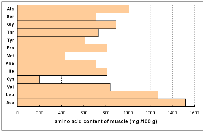
What conclusions can you draw from these results?
Although the output of alanine by muscle in fasting is 3-fold higher than any other amino acid, this does not reflect the amino acid composition of muscle proteins. Alanine accounts for ~60% of the total amino acid output from muscle, yet it accounts for only 10% of the amino acids in muscle proteins.
The diagram below shows the arterial - hepatic vein differences in concentrations of amino acids
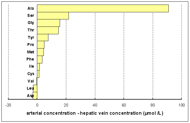
What conclusions can you draw from these results?
The liver takes up most amino acids in the fasting state (although it puts out small amounts of valine, leucine and aspartate). Again the uptake of alanine is considerably greater than that of any other amino acid, and alanine accounts for more than half the total amino acid uptake by the liver.
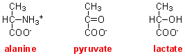 The structures
of alanine, pyruvate and lactate are shown on the right.
The structures
of alanine, pyruvate and lactate are shown on the right.
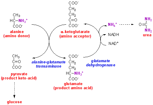
How can alanine be a substrate for gluconeogenesis?
There is a family of enzymes, transaminases, that transfer the amino group from an amino acid onto a keto-acid, so forming the amino acid corresponding to that amino acid, and leaving the keto-acid corresponding to the amino acid substrate. The keto-acid corresponding to alanine is pyruvate, which, as we have already seen, is a substrate for gluconeogenesis.
In the liver the substrate keto-acid is commonly ketoglutarate, forming glutamate. This glutamate can then undergo oxidative deamination to reform ketoglutarate and ammonium ions. The ammonium is then used for the synthesis of urea, which is excreted.
Note that although compounds such as pyruvate and ketoglutarate (and also the carbon skeletons of other amino acids) are commonly referred to as keto-acids, correctly they should be called oxo-acids. The C=O group is an oxo-group, and these compounds are not chemically ketones.
It is unfortunate that oxo-acids are more commonly called keto-acids, because this often causes confusion between these compounds and the ketone bodies (acetoacetate, hydroxybutyrate and acetone) which are formed from fatty acids in the fasting state in the liver to provide metabolic fuel for muscle and (in prolonged starvation) the brain. Elevated blood concentrations of ketone bodies leads to keto-acidosis, as seen in poorly controlled diabetes mellitus and prolonged starvation.